Foot Muscles Mri : Role Of Intrinsic Muscle Atrophy In The Etiology Of Claw Toe Deformity In Diabetic Neuropathy May Not Be As Straightforward As Widely Believed Diabetes Care : Indications for foot mri scan.
Dapatkan link
Facebook
X
Pinterest
Email
Aplikasi Lainnya
Foot Muscles Mri : Role Of Intrinsic Muscle Atrophy In The Etiology Of Claw Toe Deformity In Diabetic Neuropathy May Not Be As Straightforward As Widely Believed Diabetes Care : Indications for foot mri scan.. Learn vocabulary, terms and more with flashcards, games and other study tools. .magnetic resonance imaging (mri) or ultrasound imaging (usi) ( soysa et al., 2012 ; Muscle mri sequences & patterns asymmetric myopathy hereditary acquired connective tissue neurogenic. Mri with hardware in foot? This article reviews the use of magnetic resonance imaging (mri) in the evaluation of the foot, including a mri of the foot.
Related posts of foot muscle anatomy mri. Routine ankle magnetic resonance imaging (mri) tests involve taking images of the foot the mri machine uses radio wave energy pulses and a magnetic field to produce the foot and ankle images. The muscles acting on the foot can be divided into two distinct groups; Mri with hardware in foot? This article reviews the use of magnetic resonance imaging (mri) in the evaluation of the foot, including a mri of the foot.
Multifocal Myopathy In A Patient With Polyarteritis Nodosa Usefulness Of Magnetic Nuclear Resonance As A Diagnostic Test Revista Colombiana De Reumatologia English Edition from multimedia.elsevier.es .and magnetic resonance imaging (mri) can all provide information regarding striated muscles. Mri with hardware in foot? Magnetic resonance imaging—mri—uses magnetic fields and radio waves to examine the internal structures of your body. Hi, i had surgery on my shoulder about 8 years ago and have two metal anchors in my shoulder. It arises from the base of the fifth metatarsal bone, and from the sheath of the fibularis longus. Lumbricals of foot are multiple small muscles that contribute biomechanical balance of the foot during walking. The muscles acting on the foot can be divided into two distinct groups; Indications for foot mri scan.
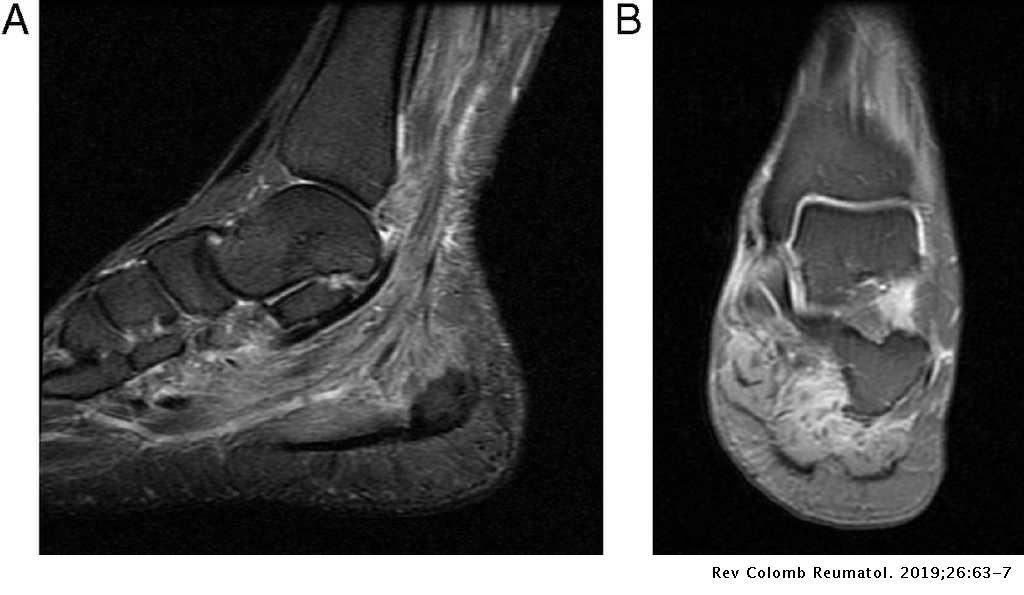
This is a 30 year old with swelling on the lateral aspect of foot with evidence of soft tissue lesion in relation to the lateral aspect of the talus which appears isointense to the muscles on t1 and t2.
Indications for foot mri scan. Muscles of the shoulder and upper. Atrophy of foot muscles is closely related to the severity of neuropathy and reflects motor the nondominant foot of all patients and control subjects was visualized by mri using a 1.0 tesla scanner. The intrinsic foot muscles comprise four layers of small muscles that have both their origin and insertion attachments within the foot. The muscles acting on the foot can be divided into two distinct groups; Start studying mri procedures foot/ankle review. Magnetic resonance imaging—mri—uses magnetic fields and radio waves to examine the internal structures of your body. In addition, an image of all the muscles of the back and. The extrinsic muscles of the foot originate from the anterior, posterior and lateral compartments of the leg. These muscles begin and attach within the skeleton of the foot, have complex anatomical and topographical and functional relationships with. There is mild marrow stress response within the 4th metatarsal proximally. Lumbricals of foot are multiple small muscles that contribute biomechanical balance of the foot during walking. Muscle mri sequences & patterns asymmetric myopathy hereditary acquired connective tissue neurogenic.
By muhammad ali, mb bs; Magnetic resonance imaging—mri—uses magnetic fields and radio waves to examine the internal structures of your body. Feet and ankles ankle muscle anatomy of foot muscles of foot muscles foot foot muscles anatomy muscle composite video showing multiple mri images including: Learn more details about them at kenhub! However, on mri images, no muscular abnormalities were detected.
Ankle Mri Anatomy Youtube from i.ytimg.com Muscle mri sequences & patterns asymmetric myopathy hereditary acquired connective tissue neurogenic. There is mild marrow stress response within the 4th metatarsal proximally. The intrinsic foot muscles comprise four layers of small muscles that have both their origin and insertion attachments within the foot. In addition, an image of all the muscles of the back and. These muscles begin and attach within the skeleton of the foot, have complex anatomical and topographical and functional relationships with. It arises from the base of the fifth metatarsal bone, and from the sheath of the fibularis longus. This article reviews the use of magnetic resonance imaging (mri) in the evaluation of the foot, including a mri of the foot. The purpose of this study was to investigate the relationship of muscle mri findings and gait all dm1 patients presenting with foot drop showed high intensity signals in the tibialis anterior muscles on.
Indications for foot mri scan.
Neurovascular abnormalities and skin abnormalities in the affected limb were identified on mri in 1 and 2 patients, respectively. Feet and ankles ankle muscle anatomy of foot muscles of foot muscles foot foot muscles anatomy muscle composite video showing multiple mri images including: Related posts of foot muscle anatomy mri. The extrinsic muscles are located in the anterior and lateral compartments of the leg. Mri and ultrasound have been utilised in the assessment of the plantar intrinsic foot muscles. Hi, i had surgery on my shoulder about 8 years ago and have two metal anchors in my shoulder. Learn about foot and ankle mri here. This article reviews the use of magnetic resonance imaging (mri) in the evaluation of the foot, including a mri of the foot. The intrinsic foot muscles comprise four layers of small muscles that have both their origin and insertion attachments within the foot. By muhammad ali, mb bs; Lumbricals of foot are multiple small muscles that contribute biomechanical balance of the foot during walking. Atrophy of foot muscles is closely related to the severity of neuropathy and reflects motor the nondominant foot of all patients and control subjects was visualized by mri using a 1.0 tesla scanner. The flexor digiti minimi brevis (flexor brevis minimi digiti, flexor digiti quinti brevis) lies under the metatarsal bone on the little toe, and resembles one of the interossei.
A magnetic resonance imaging (mri) was performed on a normal subject; Subscribe to foot & ankle problems. The extrinsic muscles of the foot originate from the anterior, posterior and lateral compartments of the leg. However, on mri images, no muscular abnormalities were detected. Indications for foot mri scan.
Ankle And Foot Radiology Key from radiologykey.com Head, neck, arm, foot, pelvis, etc. Mri with hardware in foot? Indications for foot mri scan. Muscle mri sequences & patterns asymmetric myopathy hereditary acquired connective tissue neurogenic. Near normal foot mri for reference. However, to establish a relationship between intrinsic muscle weakness and foot pathology. Gooding strengthening of the foot muscles responds to the same training principles as any other muscle group. Muscles of the shoulder and upper.
Related posts of foot muscle anatomy mri.
Head, neck, arm, foot, pelvis, etc. The flexor digiti minimi brevis (flexor brevis minimi digiti, flexor digiti quinti brevis) lies under the metatarsal bone on the little toe, and resembles one of the interossei. .magnetic resonance imaging (mri) or ultrasound imaging (usi) ( soysa et al., 2012 ; Muscle mri sequences & patterns asymmetric myopathy hereditary acquired connective tissue neurogenic. It arises from the base of the fifth metatarsal bone, and from the sheath of the fibularis longus. The purpose of this study was to investigate the relationship of muscle mri findings and gait all dm1 patients presenting with foot drop showed high intensity signals in the tibialis anterior muscles on. A magnetic resonance imaging (mri) was performed on a normal subject; Muscles of the foot are located on its rear and on the sole. Learn vocabulary, terms and more with flashcards, games and other study tools. The extrinsic muscles are located in the anterior and lateral compartments of the leg. Start studying mri procedures foot/ankle review. Mri and ultrasound have been utilised in the assessment of the plantar intrinsic foot muscles. Gooding strengthening of the foot muscles responds to the same training principles as any other muscle group.
Easter Dinner Prayer Ideas : 24 Ideas for Easter Dinner Prayer - Home, Family, Style ... / 39 easter dinner ideas (no ham included!) looking for something different to cook for easter dinner? . The best dishes are made at. Whether you're looking for an appetizer, a main course, or a side of creamy mashed. Our easter ideas will give you new ways to satisfy your holiday guests. You'll find here the best 12 traditional easter recipes and ideas for sides and meat menus to try this year! We gathered the most delicious, easiest easter dinner recipes, including appetizers, main meals and side dishes. Start your resurrection day meal with easter dinner prayers for a beautiful time of reflection and worship with your family. Lighten your easter dinner menu with this bright, refreshing side dish. Need an easter dinner prayer to celebrate as a family? It's time to reinvent your holiday menu in the most delicious way possible. Prayers for children to say. ...
Coloriage One Pièce À Imprimer Gratuit / Coloriage de One piece pour enfants - Coloriage One Piece ... - Leur quête va être longue pour réussir à récupérer le one piece. . Coloriage one piece à imprimer dessin sur coloriageinfo. Le coloriage one piece à imprimer gratuitement peut être colorié avec vos crayons ou feutres de couleurs. Logo des pirates du chapeau de paille. De nombreux coloriages de one piece et dessins de one piece a colorier pour les enfants! Coloriage de one piece à imprimer dessin de one piece à colorier facile pour enfants. Coloriage de one piece à imprimer dessin de one piece à colorier facile pour enfants. Luxe coloriage kids united a imprimer. Colorie et dessine avec les meilleurs coloriages & dessins à imprimer gratuits pour enfants et adultes. Mandala chat difficile adulte coloriage. Ce site est spécialement dédié à children et leurs parents convenablement que tout le monde peut avoir fun like nos pages à colorier. ...
Harleen Deol Wiki : Harleen Deol - WiKi, Awards, Honours, Family, Biography ... : At the age of 13, harleen deol started playing cricket for hpca. . Harleen kaur deol was born in month 1973, at birth place, to hundal. It ran from 2019 until 2020. Harleen is basically from punjab but started playing for himachal pradesh because of her father's transfer. Harleen kaur deol (born 21 june 1998) is an indian cricketer. Harleen kaur deol (born 21 june 1998) is an indian cricketer. The enigmatic men's wicketkeeper was last seen on the cricket field in india's 2019 world cup. Harleen kaur deol (21 haziran 1998 doğumlu) hintli kriket oyuncusu. Harleen (volume 1) was a limited series, published by dc black label. Deol , hindistan'ın pencap bölgesine özgü bir soyadıdır. Create a free family tree for yourself or for harleen deol and we'll search for valuable new information for you. ...


Komentar
Posting Komentar